
Annonse
Share your science:
Robots may help relieve a congested healthcare system
SHARE YOUR SCIENCE: Could you imagine healthcare institutions with robots performing ultrasound examinations?

Published
With sufficient training, robotic systems possess the
potential of achieving human capabilities in this task, offering significant
benefits to a congested healthcare system. Robotic ultrasound could lend an
extra hand in the acquisition of data, thereby augmenting the hospital's
capacity to serve more patients.
Moreover, it could standardize the examinations, facilitating easier comparisons and simplifying follow-up procedures.
Additionally, robotic ultrasound could empower a wider range of healthcare personnel to perform, or supervise, these types of examinations, thus alleviating the need for scarce experts.
Facts
- To accomplish the goals, the formation of interdisciplinary teams becomes imperative, bringing together experts with diverse backgrounds including, but not limited to, medicine, ultrasound, robotic control, and AI.
- Key members include:
Cristiana Golfetto, Andreas Østvik, Martin Albertsen Brandt, Javier Pérez de Frutos, Lars Eirik Bø, Esten Ingar Grøtli, Marialena Vagia, Klaus Ening, Geir Arne Tangen and Jan Tommy Gravdahl
Robotic ultrasound?
We at SINTEF believe in the potential of human-robot collaboration. Our goal is to develop robotic systems able to perform autonomous ultrasound examinations.
Annonse
Organs within the body are not completely stationary, they undergo movements caused by respiration. In certain medical procedures, it is crucial to know the exact position of the organ of interest.
For instance, when utilizing shock waves to disintegrate kidney stones, it is desirable to hit the stones without harming healthy tissue. Similarly, during needle insertion for biopsies, it is crucial to place and navigate the needle in the correct direction.
Advantages of robotic ultrasound
Ultrasound examinations of moving organs demand outstanding hand-eye coordination by the clinicians when looking at the screen of the scanner. Therefore, it is common practice to keep the probe still during both recordings and interventional procedures.
Robotic ultrasound platforms possess the ability to interpret images and sensor data and react and move autonomously according to what is interpreted. This enables the acquisition of stable images of moving organs, opening a range of new possibilities and enhancements.
If robots can follow moving organs, this can also unlock new measurements and monitoring opportunities, promising advancements in diagnostic capabilities.
For example, during patient follow-ups, the images are easier to compare and study over time. If robots can follow moving organs, this can also unlock new measurements and monitoring opportunities, promising advancements in diagnostic capabilities.
Artificial intelligence, ultrasound and robotic arms
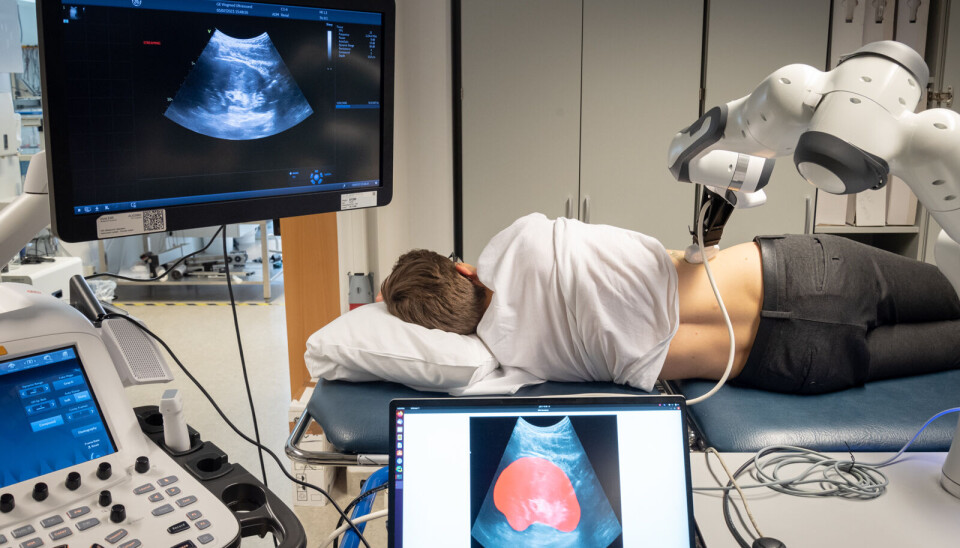
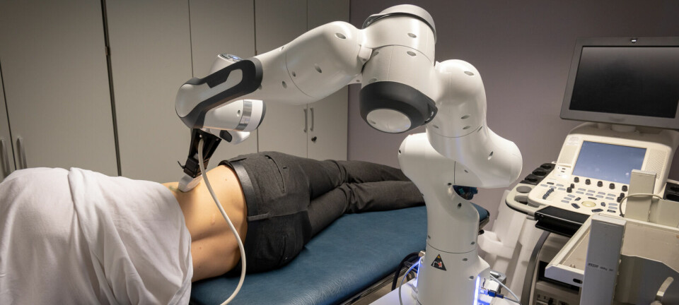
The robotic system consists of a robotic arm equipped with a US probe and an AI algorithm trained to identify the organ of interest within a US image and track its movement in real-time.
This work originates from a collaboration between researchers at SINTEF Digital and NTNU which started in 2017. The researchers explored robotic interaction with moving objects for different use cases, one of which was robotic ultrasound scanning of the abdominal aorta.
The challenging path to autonomous ultrasound scanning
There are various challenges in this project, notably the use of modern robotic arms and algorithms to engage in compliant interaction, allowing the robot to respond gently to contact, like a spring or a rubber band. This feature enhances safety when interacting with humans, but requires more sophisticated control algorithms.
It makes it especially challenging when we also need a robust algorithm for finding the organ in the ultrasound image, so the robot may follow the movement of the organ over time.
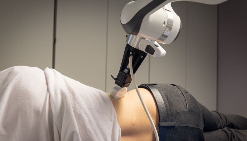
Maintaining precise control and balance throughout the entire recording process becomes paramount; we want to minimize the probe pressure to prevent patient discomfort, while simultaneously ensuring optimal acoustic contact to achieve the best possible image quality.
Undoubtedly, there are significant tasks and hurdles ahead. However, our team approaches these challenges with great enthusiasm and determination. The vision of healthcare institutions with robots working alongside humans is an inspiring goal we strive to achieve. With each step, we move closer to transforming this vision into a tangible reality.
Share your science or have an opinion in the Researchers' zone
The ScienceNorway Researchers' zone consists of opinions, blogs and popular science pieces written by researchers and scientists from or based in Norway.
Want to contribute? Send us an email!


