Annonse
New scans aid brain cancer treatment
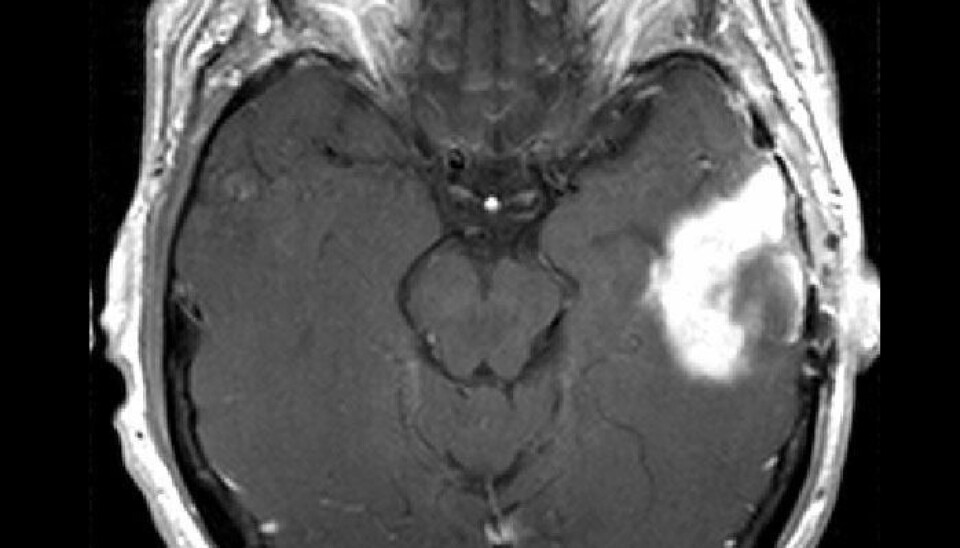
A new MRI technique offers better images of brain tumours and quicker diagnoses. It can also help doctors decide more quickly to halt the use of ineffective medications.
Denne artikkelen er over ti år gammel og kan inneholde utdatert informasjon.
This new method, called Vessel Architecture Imaging, uses an MRI scanner to get information about blood vessels in the brain.
MRI scanners use contrast agents, which act like dyes, to analyse the characteristics of a tumour's blood vessels.
Medical technicians can see how a contrast agent spreads through the blood vessels of tumours to calculate the speed of blood flow, how much blood the tissue contains and the calibre of the blood vessels.
This information enables them to doctors whether the tumour is growing and whether the patient is responding to treatment.

Information from time lags
Annonse
Medical personnel can now take two blood flow MRI recordings in parallel.
The new technique takes advantage of the small dissimilarities between the two takes, providing a much clearer picture of the changes in a tumour.
“We can observe delays between the two takes on the basis of characteristics in the tissue. The difference in these time lags provides us with new information,” explains researcher Kyrre Eeg Emblem.

Emblem was involved in the study along with a number of international researchers and colleague Professor Atle Bjørnerud at the Intervention Centre of Oslo University Hospital.
MRIs typically only measure the diameter of blood vessels, but the new method enables the identification of different types of blood vessels and their expenditure of energy in the form of oxygen intake.
Seeing whether a treatment is working
This information boosts the ability of a doctor to determine if a patient is responding to a cancer treatment regime much earlier in the game.
"We used this method on brain cancer patients who were taking tumour-inhibiting drugs. The method showed that 30 percent of the patients were responding to the treatment. These patients lived an average of six months longer than those who didn’t respond,” says Emblem.
“This new method allowed us to also spot the effect of the medication after a single day. Traditionally, we need a month or more to make this kind of determination.”
When you know that a patient is responding to a treatment or not, you can discontinue useless medication earlier in the course of the illness and thus spare the patient from wasting time on ineffective drugs and from experiencing potential bad side-effects from those drugs.
Clear for use
The new method can be also used in future treatment of vascular conditions such as strokes. The information about the blood vessels’ intake of oxygen is relevant in the assessment of damage done by a stroke and detecting areas that can be saved.
Emblem says the new method is ready for use.
“The method can be used if there is access to MRI scanners that are capable of imaging two parallel bloodstream recordings,” he explains.
Annonse
The scientists will now focus on further confirmation of their results and on investigating the method’s use on stroke victims. They will also test iton other forms of cancer, outside the brain.
-----------------------------
Read the Norwegian version of this article at forskning.no
Translated by: Glenn Ostling


