From the laboratory to the gallery: Microscopic images of our cells as art
The researchers' goal is to create artifical cells that can for instance knock out a type of cancer or neutralize harmful substances in the body. Now they are sharing their images of how life's tiny building blocks behave under the microscope.
Irep Gözen spends a lot of time studying a world that few people have ever seen.
The associate professor at the Norwegian Centre for Molecular Medicine is working to figure out how a cell is made by first looking at its most basic element: the small fat capsules where everything starts.
“Fat molecules are similar to the oil you use in your kitchen. They surround all the cells in your body and act as a barrier between the outside and the inside of the cell,” Gözen tells ScienceNorway.
The ultimate goal of Gözen and a group of researchers in Life Sciences at the University of Oslo is to be able to use cells as the inspiration to create artificial nanocapsules that can neutralize harmful substances in the environment or the body.
“We’re thinking of artificial cells that are programmed to knock out a type of cancer cell, for example,” says Gözen.
But the researcher emphasizes that that’s still a long way off. To emulate a cell, researchers first have to understand how everything works. And that’s extremely complicated.
Gözen and her research team are starting from scratch. By doing that, they also touch on new knowledge about how life might have originated.
The material is the artist
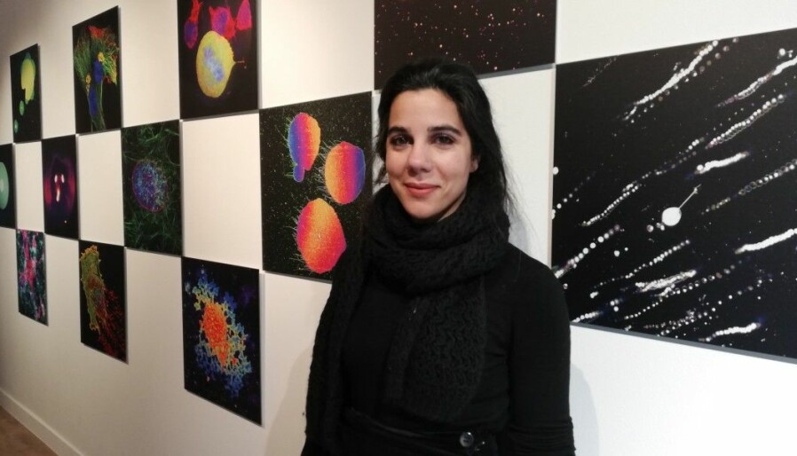
In connection with the Oslo Life Science conference in February, researchers are displaying images from the Programmable Cell-Like Compartments project that show what is going on in a micro-universe.
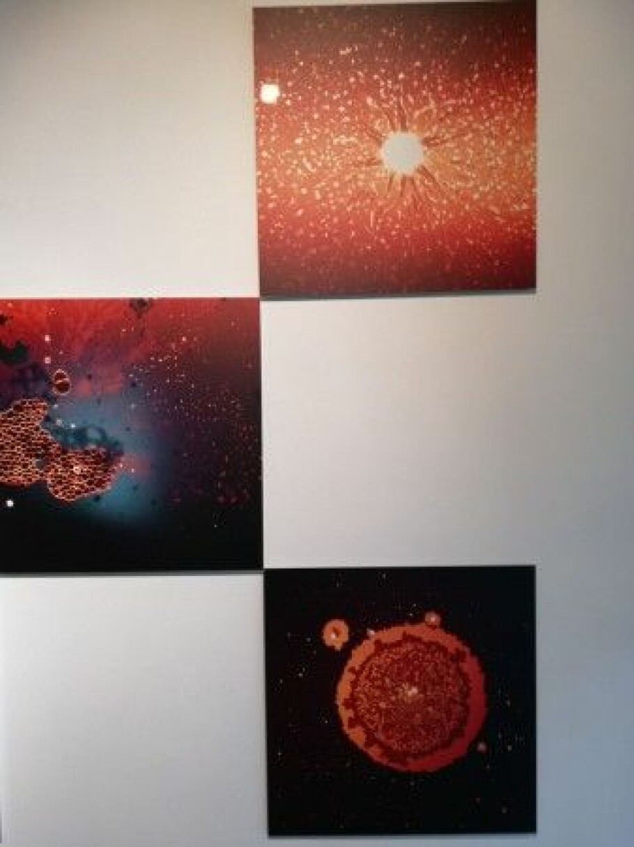
The NanoCosmos pictures are on display at Kunstplass Contemporary Art in Akersgata in Oslo. Many of the images were taken from Gözen and her group's research on lipid membranes.
“In a way, it's the material that is the artist, and we capture the moments in pictures,” says Gözen.
Some of the exhibit’s colourful images of life's micro building blocks are reminiscent of images from the macro-universe, such as stars, the sun, and nebulae.
“Pictures are everything to us – they make up our research and observations,” says the molecular biologist.
Cosmos double sense
The project team includes researchers who are working on cancer, mathematical modelling, soft materials and philosophy.
Gry Oftedal, a senior lecturer and researcher in philosophy at the University of Oslo, came up with the idea to showcase the work as art.
When she first saw the microscopic images taken by Gözen and her students, she immediately felt something more than mere intellectual curiosity.
“The pictures aren’t just incredibly beautiful. Looking at them, you know that we’re seeing something that’s been hidden from us until these highly advanced microscopes were developed,” Oftedal writes in an e-mail to ScienceNorway.
It is no coincidence that the exhibition has been named NanoCosmos. The nanoscale ranges from 1 – 100 nanometres, and the images reflect what happens on this scale that the researchers are studying. A nanometre is one billionth of a metre.
"Typically, we associate the word cosmos with the universe or space, but it can also mean an ‘orderly system’ or an ‘orderly world,’” says Oftedal.
The exhibit reflects both of these meanings of cosmos, she says.
“The nanoscale can be seen as its own ‘space,’ where researchers are working to discover the principles and regularities that apply there,” says Oftedal.
“It’s a world where materials have completely different properties than on larger scales, and at the same time we can also recognize certain patterns from the macro scale. A lot of viewers may also find that the images themselves create associations with outer space.
Stripping away everything
Gözen explains what is going on in the abstract compositions.
“We’re trying to understand what happens when you take away a cell's genetics, the mechanisms that tell the cell what to do,” she says.
The researchers strip away everything and see what the material does on its own, subject to the laws of nature.
Other researchers are investigating cells from the top down. For example, by starting with a regular cell, they remove a component and see what happens. Gözen works the other way around, starting with a basic building block, the lipid membrane, and then adding one component at a time to see if it has any effect.
“The membrane actually does a lot of interesting things on its own – without any genetics, chemical energy or proteins,” Gözen says.
The fat molecules change and move, creating different surfaces, temperatures and ions in the water. Sometimes they almost look alive.
Rupturing like an earthquake
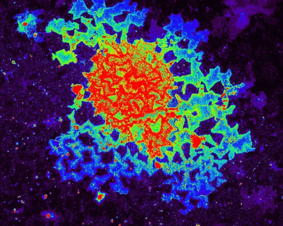
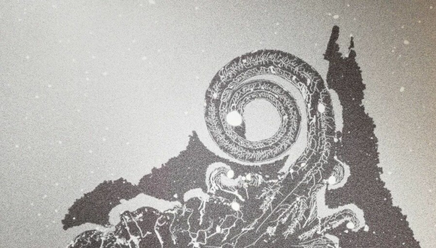
When a lipid membrane lands on a mineral surface of thin slices of stone, for example, something happens.
The membrane presses down onto the surface, which draws in the molecules. Then it starts to burst and creates patterns that look like cracks in a wall.
"Oddly enough, this rupturing activity corresponds to what happens in an earthquake,” says Gözen.
“There seems to be a universal law between how these membranes rupture and how the earth ruptures. The materials are very different, and the scales are completely different, but there seems to be a connection,” she says.
Until now, scientists haven’t understood how membranes break up. The exhibit enables us to see these microprocesses enlarged in bright imagery.
“Environmental changes can close the patterns. The pharmaceutical industry is interested in these types of mechanisms. Because if you understand all the details of how a membrane ruptures, it can give you an idea of how to programme the delivery of medicines to the cell. You have to get through the membrane to reach the cell,” says Gözen.
Hints on how the first cells were made
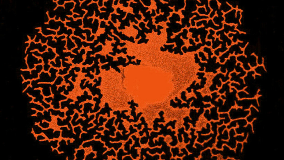
The fat capsules that Gözen has examined flatten themselves on the mineral surface, then they burst and begin to form small nanotubes. These are small cylinders 100 nanometres in diameter.
What happens next may provide clues as to how the first hereditary material on earth may have originated.
After a while, some of the tubes expand into spherical capsules that are still linked together by the nanotube web. RNA, which was probably the first hereditary material on Earth, can travel between capsules without the need for cells to divide. How cell division first began is still a mystery.
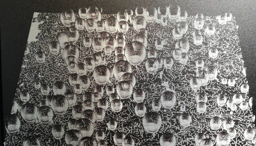
“This is a new form of cell division,” says Gözen.
The team’s research was published last year. The researchers are now investigating the phenomenon further.
By altering the presence of calcium ions, the researchers have seen how a protective capsule is formed outside the system of inner small bubbles and nanotubes.
This process is a bit similar to a cell with organelles inside, the cells organs.
“By using only fat molecules, surfaces and water, they can behave a lot like primitive cells might have done. We’re also getting clues about how today's cell systems work,” says Gözen.
Philosophical questions
As a philosopher of science, Gry Oftedal is inspired to think more about the relationship between small and large scales.
“How do we explain phenomena on different scales, and what is the relationship between these explanations? Something else that is so fascinating about the research on membranes and membrane capsules, as we can see in the pictures and videos of the exhibit, is how ‘alive’ small cell-like capsules can seem, even though they have no DNA,” she says.
The goal of the research is ultimately for capsules like these to be able to recognize harmful substances and neutralize them.
“Down the road, some of this research could also help us understand how life on Earth originated and how we might be able to create artificial cells. This raises a lot of philosophical and ethical questions in terms of how we should understand life and whether life is something we can or should try to re-create in the laboratory,” says Oftedal.
Another philosophical issue that concerns Oftedal about the project is the use of metaphors from computer science and machine reasoning in bio-nano science.
“‘Molecular machines,’ ‘nanorobots’ and ‘programmed cells’ are examples of this kind of metaphor. We’re working to understand what role these metaphors play in scientific theory and practice, and especially in scientific communication,” Oftedal says.
Translated by: Ingrid P. Nuse
———
Read the Norwegian version of this article at forskning.no


