


Annonse
How is the brain cleansed? Researchers are divided over the findings of a new Norwegian study
“It’s difficult to explain this away, because we can see it with the naked eye,” neurosurgeon Per Kristian Eide says.
Published
When two Norwegian researchers examined the brains of 75 patients, they discovered something no one had seen before.
Participants in the new study had a contrast agent injected into their spinal cords, where it mixed with the clear cerebrospinal fluid.
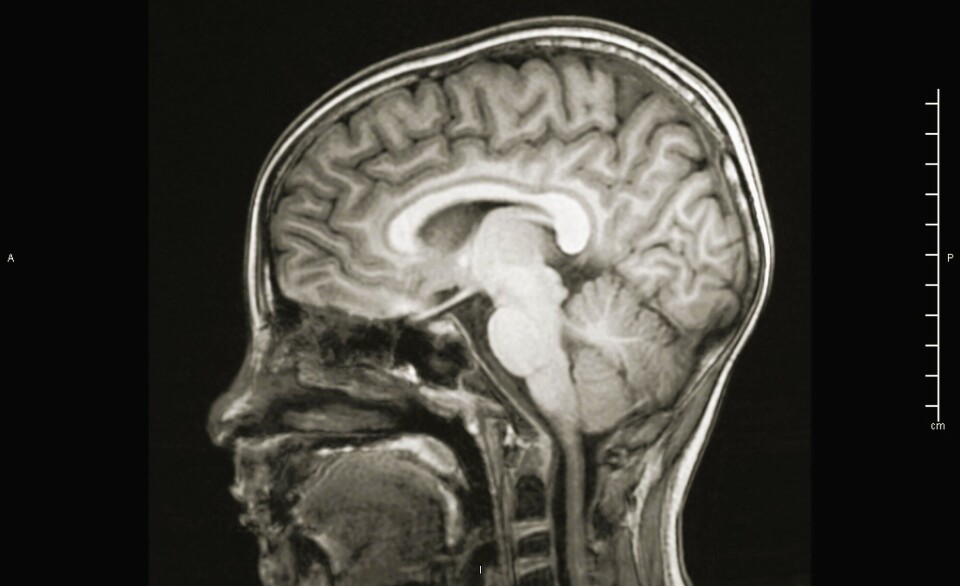
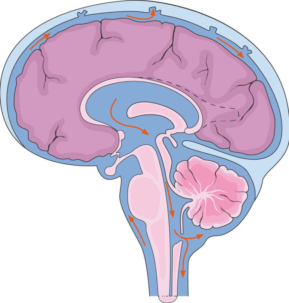
This fluid flows up to the skull, surrounds the brain, and fills the cavities.
Two to three hours after the contrast agent was injected into the spine, it lit up in a very special way on the MRI images.
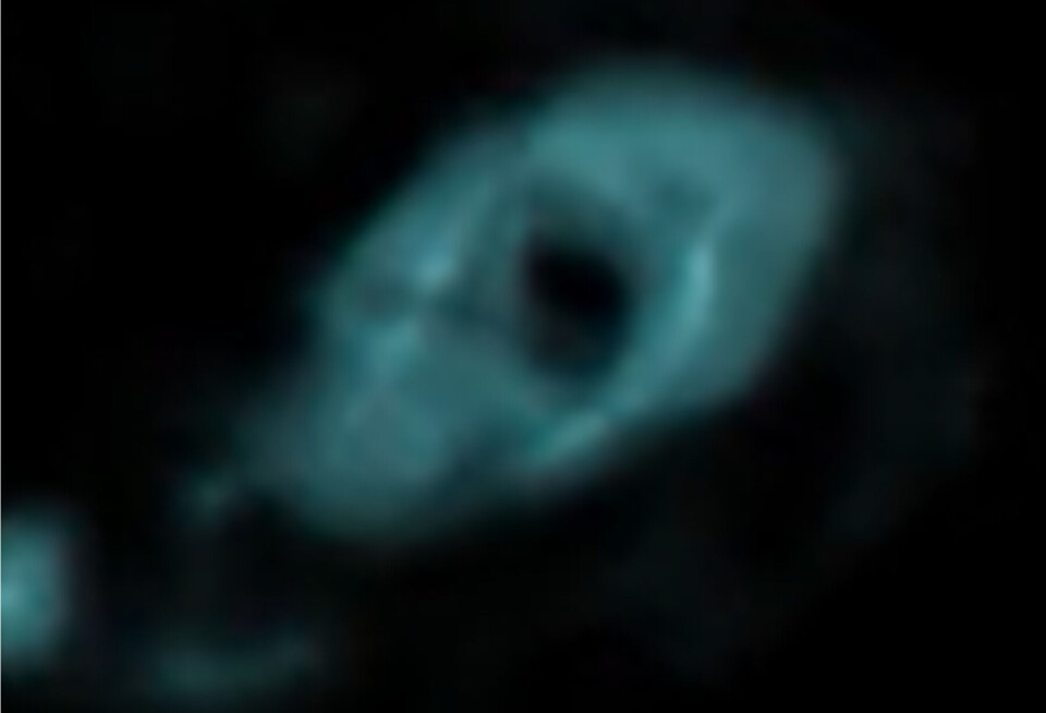
The fresh cerebrospinal fluid formed a doughnut-shaped ring around the large arteries entering the brain.
Annonse
The significance of this is at the heart of a heated dispute among neuroscientists.
The liquid that cleanses the brain
To understand the disagreement and how this glowing doughnut fits in, we must go back in time.
Cerebrospinal fluid has been underestimated for centuries, Per Kristian Eide tells sciencenorway.no. He is one of the researchers behind the new study that was published in the scientific journal Nature Communications.
Doctors have long known that the clear liquid protects the brain from impacts and regulates pressure in the skull.
But more and more research suggests that it plays another crucial role: cleansing the brain.
If this is true, the clear liquid could become a key to understanding dementia and Alzheimer's disease. Not just in what goes wrong, but also how these conditions can be detected and how they can be treated.
Divisive theories from Danish researchers
The crux of the dispute is how this brain cleansing process occurs.
“It must be that fresh cerebrospinal fluid enters and the waste exits,” Eide explains to sciencenorway.no.

But where is the evidence that this is happening?
Danish researcher Maiken Nedergaard and her colleagues have proposed several theories about how cerebrospinal fluid cleanses the brain.
Their most recent discovery, published in 2023 in the scientific journal Science, revealed the existence of a fourth brain membrane.
If this also applies to humans, textbooks will need to be rewritten.
Annonse
Since the 19th century, it has been accepted knowledge that humans have three membranes, or meninges.
Disruption in the scientific world
So, how could a fourth membrane help clean the brain?
According to classical knowledge, the fluid surrounding the brain flows around in a space between two meninges.
The fourth membrane, on the other hand, divides this fluid-filled space in two, according to Nedergaard and colleagues.
The implications of this are unclear, but the division might allow fresh cerebrospinal fluid to penetrate the brain more easily.
Other researchers are not convinced.
“This has created quite a stir in the scientific world,” says Eide.
Tubes around blood vessels
The Danish research group’s experiments were conducted on mice, highlighting the urgent need for human studies.
So, does the Norwegian study confirm that there is a fourth brain membrane encasing the entire brain?
It does not, according to the Norwegian researchers. At the same time, it cannot disprove the theory either.
The doughnut around the blood vessels shows a membrane that encapsulates some of the cerebrospinal fluid, Eide believes.
So there may be a tube-like structure surrounding the blood vessels going into the brain.
“It's difficult to explain this away, because we can see it with the naked eye. We see that cerebrospinal fluid moves along the blood vessels in the same direction as the blood, in the direction of the brain,” Eide explains.
Divided in the interpretation of the study
Fresh cerebrospinal fluid can hitch a ride with the blood vessels in this tube into the brain, the Norwegian researchers believe.

However, the disputing parties remain divided in their interpretation of the new Norwegian study.
“That is the authors' hypothesis, but it remains to be proven. I believe instead that the results could be explained much more simply,” Christer Betsholtz writes to sciencenorway.no.
Betsholtz, a neuroscientist at Karolinska Institutet, has been one of the most vocal critics of Nedergaard's theories regarding a fourth brain membrane.
Claiming that there is a fourth membrane is like saying we have three kidneys just because a new type of kidney cell has been found, he has previously told the journal The Transmitter.
Does not undermine the theory of the fourth meninge
Maiken Nedergaard at the University of Copenhagen, on the other hand, supports Per Kristian Eide and colleague Geir Ringstad's interpretation.
“It's a very good study and has significant implications for our understanding of the brain's fluid transport,” the Danish researcher writes in an email to sciencenorway.no.
She does not think it undermines her own theory of a fourth membrane surrounding the entire brain.
“Eide and Ringstad's MRI study shows that there is a membrane, but they can't say which one,” Nedergaard writes.
To truly confirm this, the tissue itself would need to be analysed, which would not have been possible since the experiments were done on living subjects, she adds.
A potential path into the brain
But despite the disagreements, all three researchers agree that there is a lot at stake.
Penetrating the brain with medications has been nearly impossible due to the strict blood-brain barrier between the blood and brain.
If a parallel stream flows into the brain, medications could be injected into the spinal cord instead.
This is already done in a few cases, but remains uncommon.
Slower flow in people with dementia
Understanding how substances are cleared from the brain is just as important.
An example is tau protein, which accumulates in the brains of people with Alzheimer's disease.
In the Norwegian study, some of the participants had dementia.
When Eide and Ringstad examined how the cerebrospinal fluid moved in this group, they saw a clear pattern: The contrast agent in the cerebrospinal fluid moved more slowly through the brain.
If cerebrospinal fluid can remove substances like tau, this could lead to new treatments.
Perhaps we can develop new drugs that increase cerebrospinal fluid flow, Nedergaard points out. This might make the cleansing process more efficient.
Alternative theory
But if we are to believe the Swedish researcher Christer Betsholtz, many unanswered questions remain before we reach that point.
Betsholtz presents an alternative theory to sciencenorway.no regarding why the glowing doughnut appears in the Norwegian study.
The MRI images may mean something entirely different than the existence of a membrane or tube for cerebrospinal fluid around the blood vessels, he believes.
Like pouring dye into a stream
The neuroscientist at Karolinska Institutet compares his explanation to running water.
Anyone who has raced toy boats in a stream knows that the water flows faster in the middle and slower along the edges.
So, if someone pours dye into the water higher up the stream, a spectator further downstream will see the colour appear in the middle of the river first.
The same can happen when the contrast agent is injected into the spine.
This means that the dyed cerebrospinal fluid reaches the blood vessels faster, which are centrally located in the fluid-filled space around the brain.
Or perhaps there is a completely different explanation, he adds.
The contrast agent reaches the entire brain
Regardless, there is no doubt that the contrast agent penetrates deep into the brain in the new study.
The Swedish brain researcher also agrees with this.
Eide and Ringstad demonstrated this back in 2015.
“At the time, when we started with this many years ago, we were very surprised because we initially believed that substances do not enter the brain. We thought they would maybe penetrate a millimetre into the brain. But no, that's not the case. It gets into the whole brain,” Eide explains.
The big mystery is still how substances in cerebrospinal fluid can enter and exit the brain and which molecules are involved, Betsholtz believes.
References:
Eide, PK & Ringstad, G. Functional analysis of the human perivascular subarachnoid space, Nature Communications, vol. 15, 2024. DOI: 10.1038/s41467-024-46329-1
Møllgård et al. A mesothelium divides the subarachnoid space into functional compartments, Science, vol. 379, 2023. DOI: 10.1126/science.adc8810
———
Translated by Nancy Bazilchuk


